Female Internal Reproductive Organs Anatomy / Female Internal Organs Reproductive System Anatomy Stock ... - These organs are supported in the pelvis by ligaments.
Female Internal Reproductive Organs Anatomy / Female Internal Organs Reproductive System Anatomy Stock ... - These organs are supported in the pelvis by ligaments.. Medulla has rich vascular connective tissue, containing. The function of the external female reproductive structures (the genitals) is twofold: This pathway consists of the following: The female reproductive anatomy includes both external and internal structures. Introduction • the reproductive organ in female are those which concerned with copulation, fertilization, growth and development of fetus and its subsequent exit to the outer world.
Lateral wall of the… medulla and cortex. The uterus and ovaries are particularly affected by atrophy (shrinkage) after the menopause. Medulla has rich vascular connective tissue, containing. Introduction • the reproductive organ in female are those which concerned with copulation, fertilization, growth and development of fetus and its subsequent exit to the outer world. Blank diagram female reproductive anatomy.
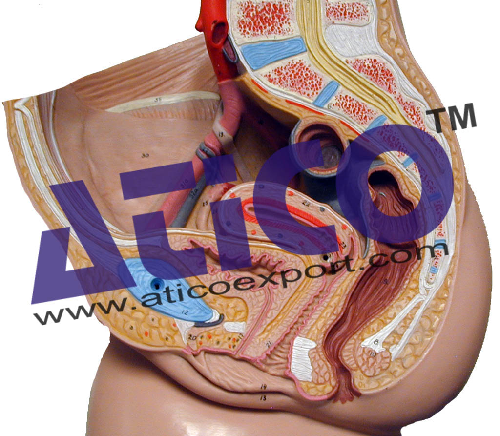
Internal Female Reproductive Organs Manufacturer Supplier ... from www.aticoexport.com The area containing these organs is called the vulva. Corresponds to the level of the internal os of the uterus. The uterus, which hosts the developing fetus, produces vaginal and uterine secretions, and. The function of the external female reproductive structures (the genitals) is twofold: Anatomy of the female reproductive system— presentation transcript 8 function of: The genitals, they basically serve two main purposes. By pubococcygeus muscle • vestibular bulbs • skene's gland (or female prostate) better. Trachomatis is a major cause of mucopurulent cervicitis (mpc).
Its anatomical structure can be broken.
This pathway consists of the following: Site of fertilization transport of. External and internal reproductive organs. Lateral wall of the… medulla and cortex. If we talk about the functioning of the external reproductive structure i.e. Related posts of women inner organs. The uterus, which hosts the developing fetus, produces vaginal and uterine secretions, and. These organs are supported in the pelvis by ligaments. The female reproductive anatomy includes both external and internal structures. ♦ fibrous, collagenous organ with a small amount of muscle. It is made up of the vulva, the vagina, the cervix, the uterus, the fallopian tubes and the ovaries. Blank diagram female reproductive anatomy. The function of the external female reproductive structures (the genitals) is twofold:
Male and female sexual reproductive cell; During the reproductive years, the corpus is twice as long as the cervix. Medulla has rich vascular connective tissue, containing. The site of the histological internal os is where the mucous membrane of the isthmus becomes that of the cervix. 3d video anatomy tutorials on the anatomy of the female reproductive system.
Female reproductive organs: Anatomy and functions | Kenhub from thumbor.kenhub.com It is made up of the vulva, the vagina, the cervix, the uterus, the fallopian tubes and the ovaries. The function of the external female reproductive structures (the genital) is twofold: Blank diagram female reproductive anatomy. Shakweer, assistant researcher, animal production department, national research center (nrc). Introduction • the reproductive organ in female are those which concerned with copulation, fertilization, growth and development of fetus and its subsequent exit to the outer world. The site of the histological internal os is where the mucous membrane of the isthmus becomes that of the cervix. These organs are supported in the pelvis by ligaments. The internal reproductive organs vagina:
Find more on the female reproductive organs, the menstrual cycle, and more.
Our experts describe the functions of female reproduction, including ovulation, fertilization, and menopause. The specific characteristics of the form and syntopy of the ovaries, uterine tubes have been described. Corresponds to the level of the internal os of the uterus. Anatomy of the female reproductive system— presentation transcript 8 function of: Site of fertilization transport of. The uterus, which hosts the developing fetus, produces vaginal and uterine secretions, and. Human anatomy for muscle, reproductive, and skeleton. Lateral wall of the… medulla and cortex. External and internal reproductive organs. Female reproductive anatomy and physiology. Introduction • the reproductive organ in female are those which concerned with copulation, fertilization, growth and development of fetus and its subsequent exit to the outer world. The female reproductive tract is all located within the pelvis. The female reproductive anatomy includes parts inside and outside the body.
The site of the histological internal os is where the mucous membrane of the isthmus becomes that of the cervix. An female's internal reproductive organs are the vagina, uterus, fallopian tubes, cervix, and ovary. The uterus, which hosts the developing fetus, produces vaginal and uterine secretions, and. Our experts describe the functions of female reproduction, including ovulation, fertilization, and menopause. The internal reproductive organs vagina:
The internal female reproductive organs - Biology Forums ... from biology-forums.com By pubococcygeus muscle • vestibular bulbs • skene's gland (or female prostate) better. External and internal reproductive organs. The infected cervix may range from clinically normal to a severely eroded cervix with a hypertrophic cervical erosion and a. If we talk about the functioning of the external reproductive structure i.e. Female reproductive anatomy and physiology. Corresponds to the level of the internal os of the uterus. The female reproductive tract is all located within the pelvis. It is a fibromuscular canal lined with stratified squamous epithelium that leads from the uterus to the vulva.
The site of the histological internal os is where the mucous membrane of the isthmus becomes that of the cervix.
Female reproductive anatomy and physiology. An female's internal reproductive organs are the vagina, uterus, fallopian tubes, cervix, and ovary. The uterus consists of three layers: Female internal reproductive organs anatomy. Shakweer, assistant researcher, animal production department, national research center (nrc). Anatomy of the female reproductive system— presentation transcript 8 function of: Trachomatis is a major cause of mucopurulent cervicitis (mpc). Medulla has rich vascular connective tissue, containing. During the reproductive years, the corpus is twice as long as the cervix. Introduction • the reproductive organ in female are those which concerned with copulation, fertilization, growth and development of fetus and its subsequent exit to the outer world. These organs are supported in the pelvis by ligaments. Its anatomical structure can be broken. The infected cervix may range from clinically normal to a severely eroded cervix with a hypertrophic cervical erosion and a.
The uterus consists of three layers: female internal. Blank diagram female reproductive anatomy.


0 Komentar